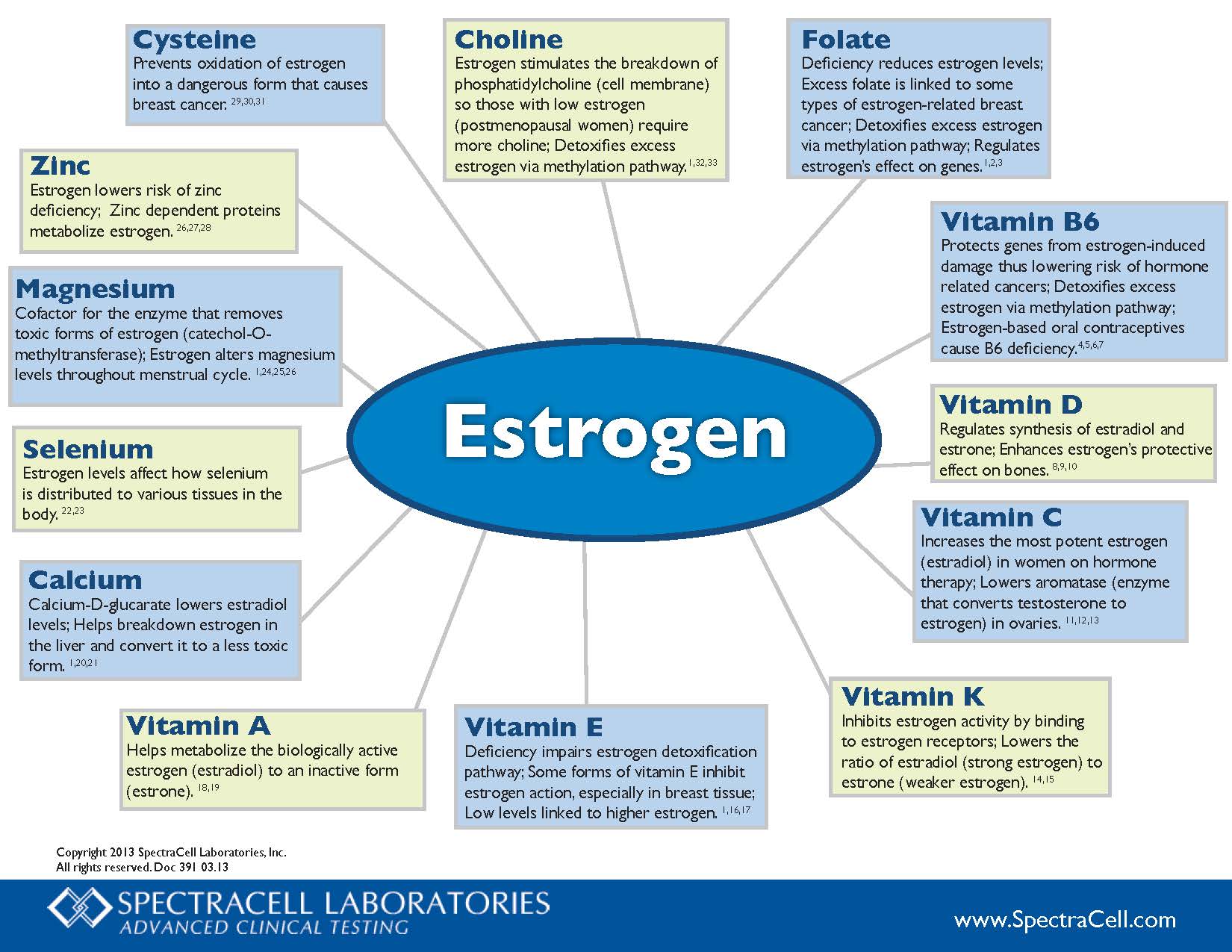
Choline – Estrogen stimulates the breakdown of phosphatidylcholine (cell membrane) so those with low estrogen (postmenopausal women) require more choline; Detoxifies excess estrogen via methylation pathway.1,32,33

Folate –  Deficiency reduces estrogen levels; Excess folate is linked to some types of estrogen-related breast cancer; Detoxifies excess estrogen via methylation pathway; Regulates estrogen’s effect on genes.1,2,3

Vitamin B6 – Protects genes from estrogen-induced damage thus lowering risk of hormone related cancers; Detoxifies excess estrogen via methylation pathway; Estrogen-based oral contraceptives cause B6 deficiency.4,5,6,7

Vitamin D – Regulates synthesis of estradiol and estrone; Enhances estrogen’s protective effect on bones.8,9,10

Vitamin C – Increases the most potent estrogen (estradiol) in women on hormone therapy; Lowers aromatase (enzyme that converts testosterone to estrogen) in ovaries.11.12.13

Vitamin K – Inhibits estrogen activity by binding to estrogen receptors; Lowers the ratio of estradiol (strong estrogen) to estrone (weaker estrogen).14,15

Vitamin E – Deficiency impairs estrogen detoxification pathway; Some forms of vitamin E inhibit estrogen action, especially in breast tissue; Low levels linked to higher estrogen.1,16,17

Vitamin A – Helps metabolize the biologically active estrogen (estradiol) to an inactive form (estrone).18,19

Calcium –  Calcium-D-glucarate lowers estradiol levels; Helps breakdown estrogen in the liver and convert it to a less toxic form.1,20,21

Selenium – Estrogen levels affect how selenium is distributed to various tissues in the body.22,23

Magnesium – Cofactor for the enzyme that removes toxic forms of estrogen (catechol-O-methyltransferase); Estrogen alters magnesium levels throughout menstrual cycle.1,24,25,26

Zinc – Estrogen lowers risk of zinc deficiency; Zinc dependent proteins metabolize estrogen.26,27,28

Cysteine –  Prevents oxidation of estrogen into a dangerous form that causes breast cancer.29,30,31

Low estrogen levels can make women more vulnerable to trauma at some points in their menstrual cycles, while high levels of the female sex hormone can partially protect them from emotional disturbance, according to new research.

…Low estrogen levels can make women more vulnerable to trauma at some points in their menstrual cycles, while high levels of the female sex hormone can partially protect them from emotional disturbance, the research suggests. 

Depression and anxiety disorders are twice as common in women as in men, but the reason for this gender difference is unclear. The new work, reviewed by Harvard’s Mohammed Milad and colleagues in a commentary, suggests that women are most at risk for symptoms of post-traumatic stress disorder (PTSD) when their estrogen is low during the menstrual cycle.

…Estrogen calms the fear response in healthy women and female rats, according to the Harvard researchers, who were led by Kelimer Lebron-Milad, an HMS instructor of psychiatry. The Emory researchers, led by postdoctoral researcher Ebony Glover, showed that the same is true for women suffering from PTSD. The higher the estrogen was in their blood when they trained on a fear-extinction task, the less likely women were to startle.

…PTSD is common in women after a trauma such as rape or sexual assault, which studies say are experienced by 25 to 30 percent of women in their lifetimes, and the symptoms last on average four times as long in women as in men after trauma. This new research suggests the reason for this susceptibility may be the monthly menstrual change in estrogen.

Steroid hormones regulate metabolic flexibility at the level of the mitochondria. Estradiol, the most frequently studied among the steroid hormones, plays a pivotal role in determining how food fuel is converted into cellular fuel or ATP. 

When we eliminate estradiol with medications such Lupron and other GnRH agonists or antagonists, or when we remove a woman’s ovaries, depleting her primary source for estrogen synthesis, metabolic flexibility diminishes significantly.*  

With the lack of metabolic flexibility comes a number of health issues, some noticeable, like weight gain, and others less noticeable, at least initially, like cardiac and neurodegenerative diseases.

READ MORE

/

Catechol-O-Methyltransferase (COMT) is one of the several enzymes that degrade dopamine, epinephrine, and norepinephrine. COMT breaks down dopamine mostly in the part of the brain responsible for higher cognitive or executive function (prefrontal cortex).

COMT helps break down estrogen byproducts that have the potential to cause DNA mutations and cause cancer. 

If you have higher COMT levels:

  • Mucuna  to increase dopamine,
  • Tyrosine to increase dopamine,
  • EGCG/Tea (COMT inhibitor),
  • Epicatechins/Chocolate (COMT inhibitor),
  • Luteolin 

If you have lower levels of COMT, the following may counteract some of the effects of the gene:

  • SAM-e â€“ however, this can increase dopamine levels in people who already have high dopamine.
  • Methyl Guard Plus to ensure adequate B6, B12, folate and betaine to support the formation of S-adenosylmethionine and prevent elevated homocysteine; S-adenosylhomocysteine inhibits COMT activity.
  • Ensure adequate anti-oxidants to prevent oxidation of dopamine and pro-carcinogenic 4-hydroxyestrogens,
  • Magnesium Citrate (magnesium is a cofactor)
  • Be careful of the following supplements that are the targets of COMT: quercetin, rutin, luteolin, EGCG, catechins, Epicatechins, Fisetin, Ferulic acid, Hydroxytyrosol
  • Avoid excessive alcohol consumption.  Since alcohol-induced euphoria is associated with the rapid release of dopamine in limbic areas, low activity COMT variant would have a relatively low dopamine inactivation rate, and therefore would be more vulnerable to the development of alcohol dependence.
  • Avoid stimulants, especially amphetamines.  Amphetamines may do worse with people who are AA, but later studies did not replicate this.  It could be differences in study design.
  • Avoid chronic stress (stress hormones require COMT for degradation and compete with estrogens),

Catechol Estrogens, Cancer and Autoimmunity

Catechol estrogens form from CYP enzymes breaking down Estradiol and Estrones. Catechol estrogens can break DNA and cause cancer and autoimmune conditions. COMT methylates (using SAM) and inactivates these catechol estrogens (2- and 4-hydroxycatechols). The products of COMT methylation are 2- and 4-o-methylethers, which are less harmful and excreted in the urine (they have anti-estrogen properties). However, if COMT is inhibited too much either because of genetics or dietary inhibition, it should result in higher levels of catechol estrogens, especially if glucuronidation and sulphation pathways are not working. 4-Hydroxyestrone/estradiol was found to be carcinogenic in the male Syrian golden hamster kidney tumour model, whereas 2-hydroxylated metabolites were without activity. 4-Hydroxyestrogen can be oxidized to quinone intermediates that react with purine base of DNA, resulting in depurination adduct that generates cancerous mutations. Quinones derived from 2-hydroxyestrogens are less toxic to our DNA. Estrone and estradiol are oxidized to a lesser amount to 2-hydroxycatechols by CYP3A4 in the liver and by CYP1A in extrahepatic tissues or to 4-hydroxycatechols by CYP1B1 in extrahepatic sites, with the 2-hydroxycatechol being formed to a larger extent .

It has been observed that tissue concentration of 4-hydroxyestradiol is highest in malignant cancer tissue, out of all the estrogens. The concentration of these Catechol Estrogens in the hypothalamus and pituitary are at least ten times higher than parent estrogens. Catechol Estrogens have potent endocrine effects and play an important role in hormonal regulation (those produced by hypothalamus and pituitary).

Increased availability of estrogen and estradiol for binding and hypothalamic sites would facilitate the formation of Catechol Estrogens. These estrogens affect Luteinizing Hormone (LH) and maybe follicle-stimulating hormone (FSH) and prolactin. Catecholestradiol competes with estradiol for estrogen binding sites in the anterior pituitary gland and hypothalamus and dopamine binding sites on anterior pituitary membranes.

Other possible mechanisms of inactivation of these catechol estrogens include conjugation by glucuronidation and sulphation. High concentration of 4-hydroxylated metabolites caused insufficient production of methyl, glucuronide or sulfate conjugate which in turn results in catechol estrogen toxicity in cells and oxidation to semiquinone and quinone, which may reduce glutathione (GSH). These oxidation products could lead to DNA mutations. The quinone/semiquinone redox system produces superoxide ions (O2ÂŻ ) which can react with NO to form peroxynitrite, which could cause DNA damage. In summary, CEs lead to the production of potent ROS, capable of causing DNA damage, thus playing an important role not only in causing cancer but also in systemic lupus erythematosus (SLE) and Rheumatoid Arthritis. The abilities of the estrogens to induce DNA mutations were ranked as follows: 4-hydroxyestrone (most damaging) > 2-hydroxyestrone > 4-hydroxyestradiol >2-hydroxyestradiol > > Estradiol, Estrone.

Fibroids – uterine leiomyomas (ULMs) – are estrogen-dependent tumors that are more common in African American women. The aetiology for such ethnic disparity is currently unknown. Catechol-O-methyltransferase (COMT) is an essential enzyme in estrogen metabolism.

Women with the high-activity COMT genotype are 2.5 times more likely to develop ULMs than women with other genotypes. The prevalence of this genotype was significantly higher in African American women (47%) compared with white (19%) or Hispanic (30%) women. Myometrial cell lines expressing the Val/Val genotype exhibited significantly enhanced responses to estrogen in proliferation and in estrogen-responsive element reporter assays. COMT-specific inhibitors reversed such a response and induced apoptosis. Myometrial specimens from Val/Val women demonstrated distinct estrogen-regulated gene expression that was consistent with enhanced proliferation and decreased apoptosis.

The results provide a possible explanation for the higher prevalence of ULMs among African American women and offer a potential new target for nonsurgical treatment using COMT inhibitors.

Postmenopausal women with a more diverse population of gut bacteria may be more efficient at breaking down estrogen, a new study suggests. Because estrogen plays a role in causing breast cancer, researchers speculate a healthy bacterial population may lower the risk for cancer.

“The composition and diversity of the intestinal microbiota were associated with patterns of estrogen metabolism that are predictive of the risk of breast cancer in postmenopausal women,”

Estrogen is metabolized in the liver and in other tissues such as the breast, yielding fragments that are excreted in urine or, through bile, into the gut. Gut microbes can degrade these metabolites, allowing them to be reabsorbed into the bloodstream and further recycled in the liver. Dr. Fuhrman and colleagues suggest that women whose gut bacteria more efficiently process estrogen may have a lowered risk for breast cancer.

Get notified about new editions

Subscribe to the Sunday Supplement

Connecting women, science and spirit, the Gynelogic Sunday Supplement delivers a bi-monthly dose of  news, views and reviews, as seen through my lady lens.