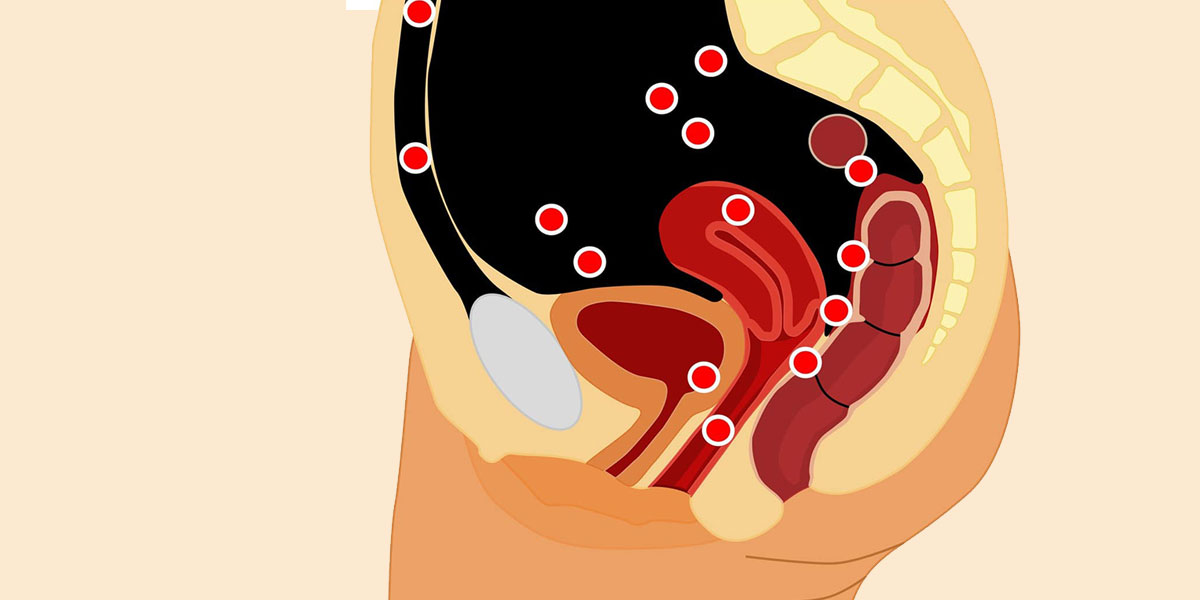
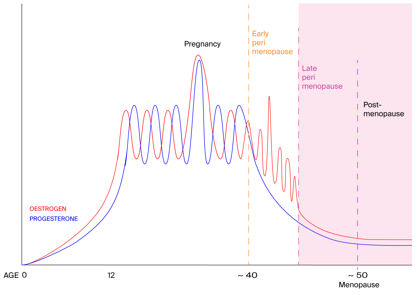
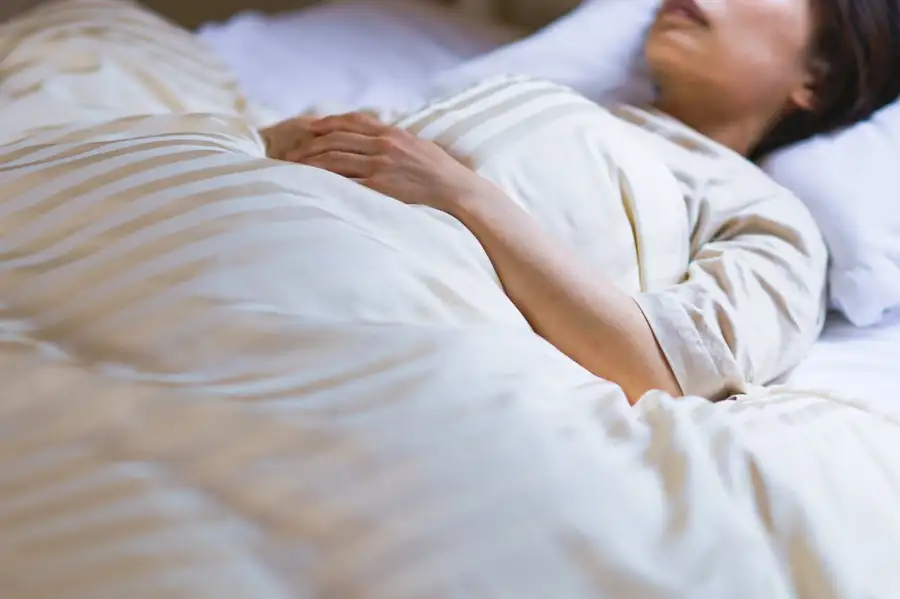
Reduced levels of oestrogen and progesterone seem to be what makes post-menopausal women more likely to have symptoms of sleep apnoea, including snoring, irregular breathing or gasping at night.

Middle-aged women who have lower levels of oestrogen and progesterone are more likely to snore, breathe irregularly and gasp while sleeping, which are all symptoms of sleep apnoea.

The involvement of these chemicals means targeted hormone therapy might prove useful for post-menopausal women, says Kai Triebner at the University of Bergen in Norway.

“Women live, on average, longer than men, but during later years, the quality of women’s life is comparatively low, which is inherently associated with their [low-oestrogen] hormone profile,” says Triebner. “Snoring and sleep-related breathing problems add to the burden.”

His team interviewed 774 women aged between 40 and 67 years old, mostly white, living in seven European countries about their respiratory health and lifestyles. The team also carried out clinical exams and took blood samples. The women, some of whom hadn’t yet reached menopause, completed questionnaires about their sleep habits and health. The study didn’t include pre or post-menopausal trans men.

Nearly half the women reported that they had a “disturbing snore”, says Triebner. In addition, 14 per cent had irregular breathing and 13 per cent gasped while sleeping.

Blood analyses revealed that the participants’ oestrogen and progesterone levels varied widely, ranging from just a few units per litre in some women to tens of thousands of units per litre in others. Those variations had clear associations with sleep apnoea, he says. As the levels of oestrone – a kind of oestrogen – doubled, women were 19 per cent less likely to snore. And as progesterone levels doubled, women were 9 per cent less likely to snore.

Within the group of women who snored, there was a 20 per cent drop in the chances of having irregular breathing as oestrogen levels doubled. And a doubling in progesterone levels was linked to a 12 per cent lower likelihood of waking up feeling like they are choking.

PLoS One DOI: 10.1371/journal.pone.0269569

The UK’s “unresponsive and defensive” healthcare system has failed thousands of women who developed life-changing conditions after pelvic mesh surgery, according to a review into the treatment.

“The report is hard hitting, harrowing and recognises the total failure in patient safety, regulation and oversight in the UK,” Kath Sansom of campaign group Sling the Mesh said in a statement.

The Independent Medicines and Medical Devices Safety Review, led by Julia Cumberlege and announced by then-health secretary Jeremy Hunt in 2018, has involved two years of data gathering from women who received vaginal and other pelvic mesh implants, mostly to treat stress urinary incontinence and prolapse that developed after childbirth.

Many women went on to develop chronic pain, nerve damage, bowel conditions, recurring infections and mobility issues, among others. The mesh can become embedded in surrounding tissues, making it very difficult to remove. The review found that it is unclear whether the mesh can change in shape or size after it is implanted, and whether chemicals from the mesh can trigger immune conditions, which have been experienced by some women.

The number of women affected by these complications is unknown, but thousands have joined support and campaign groups. Many women weren’t told about the risks of the procedure, and describe how their symptoms and complaints were dismissed by doctors as normal consequences of childbirth or menopause, says the review.

“The narrative is common,” says Sohier Elneil, a urogynaecologist and uroneurologist in London, who says she comes across similar cases on a daily basis. “Patient safety must be key to everything we do,” she says. “It should be a given, but, quite clearly from the report, it hasn’t been.”

People who have been pregnant or have breastfed a baby are less likely to experience an early menopause. This may be because ovulation is temporarily stopped during pregnancy and slowed down during breastfeeding, maintaining a reserve of eggs for longer.

The team found that people who had experienced pregnancies that lasted at least six months had a lower risk of experiencing an early menopause – defined as menopause before the age of 45 – than those who hadn’t.

“We observed a linear trend,” says Langton. “Women who had one pregnancy had an 8 per cent lower risk, those who had two pregnancies had a 16 per cent lower risk, and those that had three pregnancies had a 22 per cent lower risk.”

The link isn’t explained by infertility, says Langton. Her team accounted for this by removing people who had reported that they were trying to conceive but hadn’t become pregnant from the study sample. “There was no difference in the results,” says Langton.

Breastfeeding also lowered the risk of early menopause. People who breastfed for a total of seven to 12 months over their lifetime who had any number of infants were 28 per cent less likely to experience menopause before the age of 45 than those who had breastfed for less than a month. Those who exclusively breastfed for a total of seven to 12 months over their lifetime and who had three pregnancies had a 32 per cent lower risk of early menopause.

JAMA Network Open DOI: 10.1001/jamanetworkopen.2019.19615

Social stress may release hormones that affect bone loss, a finding that might be linked to the higher incidence of bone fractures after the menopause.

In a study of more than 8000 women aged 50 to 79, researchers found that those who reported higher levels of social stress – defined as strained relationships or stress related to social ties – were also at higher risk of bone fractures.

Women who reported high social strain and poorer quality relationships – and therefore, higher levels of stress – were found to have a larger decline in their bone density measurements over these years.

After adjusting for age, race, education, and other life style effects such as smoking and hormone therapy use, the team found that for each point of higher social strain as measured by the questionnaires, there was an associated increase of about 0.08 per cent loss of bone mineral density at the femoral neck – a portion of the hip. They also saw about 0.1 per cent greater loss across the whole hip, and about 0.7 per cent greater loss at the lower spine.

Previous research found that higher levels of stress hormones such as cortisol were associated with lower bone mineral density in the spine, and the team suggests that social stress may increase fracture risk by altering bone-regulating hormones.

Postmenopausal women may be more likely to experience social stress than their male peers. “There is research showing that social stress is higher in aging women than in men and this may be attributed to women being more likely to be caregivers in older age,” says Follis.

The team found that women with low social strain tended to be more educated and more physically active than those with high social strain. Black, Latina, and Native American women were more likely to report high social strain than White and Asian women.

Adenomyosis is a common gynecological disorder traditionally viewed as “elusive”. Several excellent review papers have been published fairly recently on its pathogenesis, and several theories have been proposed. However, the falsifiability, explanatory power, and predictivity of these theories are often overlooked. Since adenomyosis can occur spontaneously in rodents and many other species, the animal models may help us unveil the pathogenesis of adenomyosis. This review critically tallies experimentally induced models published so far, with a particular focus on their relevance to epidemiological findings, their possible mechanisms of action, and their explanatory and predictive power.

PubMed was exhaustively searched using the phrase “adenomyosis and animal model”, “adenomyosis and experimental model”, “adenomyosis and mouse”, and “adenomyosis and rat”, and the resultant papers were retrieved, carefully read, and the resultant information distilled. All the retrieved papers were then reviewed in a narrative manner.

Among all published animal models of adenomyosis, the mouse model of adenomyosis induced by endometrial–myometrial interface disruption (EMID) seems to satisfy the requirements of falsifiability and has the predictive capability and also Hill’s causality criteria. Other theories only partially satisfy Hill’s criteria of causality. In particular, animal models of adenomyosis induced by hyperestrogenism, hyperprolactinemia, or long-term exposure to progestogens without much epidemiological documentation and adenomyosis is usually not the exclusive uterine pathology consequent to those induction procedures. Regardless, uterine disruption appears to be a necessary but not sufficient condition for causing adenomyosis.

EMID is, however, unlikely the sole cause for adenomyosis. Future studies, including animal studies, are warranted to understand how and why in utero and/or prenatal exposure to elevated levels of estrogen or estrogenic compounds increases the risk of developing adenomyosis in adulthood, to elucidate whether prolactin plays any role in its pathogenesis, and to identify sufficient condition(s) that cause adenomyosis.

Background: Vitamin E is well known for its antioxidant property and has potential role in treatment of infertility. Fluoxetine is an antidepressant from SSRI group having effect on reproductive organs by increasing oxidative stress.
Aim: To explore the role of vitamin E on uterine myometrium after treatment with fluoxetine

Methods: 8-12 weeks old female rats albino of wistar strain (n=10) were divided into three groups Group A (control), group B (experimental) and group C (protective) and were treated with distilled water, oral fluoxetine (80mg/kg) and oral fluoxetine along with vitamin E simultaneously (80mg/kg and 250mg/kg) respectively for 15 days. Gross and histological examination of uterine horns of all rats along with measurement of serum prolactin level was done on 15th day. Mean±SD, One-way ANOVA and fisher exact test was applied to analyze data

Results: Weight, volume of uterus, utero-somatic index, serum prolactin level and myometrial thickness was significantly increased (P value=0.001) along with prescence of adenomyosis in experimental group B which was given fluoxetine alone. however its was observed that in group C which was given vitamin E along with fluoxetine, ameliorated these changes and results were comparable with control group A.

Conclusion: Vitamin E has a role in protection of reproductive organs owing to its antioxidant properties when administered along with fluoxetine which produces oxidative stress related changes serum prolactin levels and uterine myometrium.

Female reproductive organs have a delicate relationship with hormonal levels. Savaskan et al., 2007 observed fluoxetine use related oxidative stress and commented on its role in alteration of histological structure of reproductive organs. Jan et al., 2008 reported hormonal imbalance in rats upon treated with fluoxetine. Mori et al., 1999 observed changes in ultra-structure of uterus caused by increase in serum prolactin levels.

The animals of group C when treated simultaneously with fluoxetine along with vitamin E, showed normal thickness and histological architecture of myometrium which is similar to findings in animals of control group A. Vitamin E has been reported to ameliorate the hormonal changes inflicted by raised level of oxidative stress in body (Yin et al., 2012) .A study done by Jalili et al. 2014 supports the observations made in current study on protective ability of vitamin E in reproductive organs because of its antioxidant properties.


Medication use is a common cause of hyperprolactinemia, and it is important to differentiate this cause from pathologic causes, such as prolactinomas.

To ascertain the frequency of this clinical problem and to develop treatment guidelines, the medical literature was searched by using PubMed and the reference lists of other articles dealing with hyperprolactinemia due to specific types of medications.

The medications that most commonly cause hyperprolactinemia are antipsychotic agents; however, some newer atypical antipsychotics do not cause this condition.

Other classes of medications that cause hyperprolactinemia include antidepressants, antihypertensive agents, and drugs that increase bowel motility.

Hyperprolactinemia caused by medications is commonly symptomatic, causing galactorrhea, menstrual disturbance, and impotence.

It is Important to ensure that hyperprolactinemia in an Individual patient is due to medication and not to a structural lesion in the hypothalamic/pituitary area; this can be accomplished by (1) stopping the medication temporarily to determine whether prolactin levels return to normal, (2) switching to a medication that does not cause hyperprolactinemia (in consultation with the patient’s psychiatrist for psychoactive medications), or (3) performing magnetic resonance imaging or computed tomography of the hypothalamic/pituitary area. If the patient’s hyperprolactinemia is symptomatic, treatment strategies include switching to an alternative medication that does not cause hyperprolactinemia, using estrogen or testosterone replacement, or, rarely, cautiously adding a dopamine agonist.

Medications That May Cause Hyperprolactinemia

  • Antidepressants
    • Tricyclic and tetracyclic antidepressants
    • Monoamine oxidase inhibitors
    • Selective serotonin reuptake inhibitors
    • Other
  • Opiates and cocaine
  • Antihypertensive medications
    • Verapamil
    • Methyldopa
    • Reserpine
  • Gastrointestinal medications
    • Metoclopramide
    • Domperidone
    • Histamine2 receptor blockers?
  • Protease inhibitors?
  • Estrogens

Introduction: Fluoxetine is a commonly prescribed drug which is used in the psychiatric practice and adenomyosis is a common medical problem in women of the reproductive age group.

Objective: To explore the role of fluoxetine in the causation of adenomyosis.

Methods: Female Wistar rats (n=18) were divided into three groups (group I (the control), group II and group III) and they were treated with normal saline and oral fluoxetine (4mg/kg and 8 mg/kg) respectively for 100 days. Periodic serum prolactin measurements and histopathological examinations of the uterine horns of all the rats were done at the end. Comparison of the mean serum prolactin levels between the patients (n=15) who were diagnosed with adenomyosis, the healthy age sex matched controls and the female patients (n=20) who received fluoxetine for more than 3 months, before and after the fluoxetine administration, was done separately. Appropriate (paired or unpaired) t tests were used for the data analysis.

Results: Out of the 12 test group rats, 10 rats showed the features of adenomyosis histopathologically, along with significantly (p < 0.05) raised serum prolactin levels. The mean serum prolactin levels of the patients of adenomyosis in comparison to those of the controls and of the patients who were treated with fluoxetine (before and after the fluoxetine administration), were significantly high (p=0.001 in both the cases).

Conclusion: Fluoxetine may have some role in the causation of adenomyosis (via raising serum prolactin levels); although for a stronger evidence, the follow-up of the patients who are treated with fluoxetine on a long term basis should be ideal.

Objective: To identify clinical, laboratory and molecular genetic predictors of menstrual circle regulation in patients with polycystic ovary syndrome (PCOS) undergoing metformin treatment.

Materials and methods: The study included 143 women with PCOS (mean age is 26.4±4.6 years, mean body mass index is 23.8 (4.8) kg/m2). The assessment of androgen profile and levels of AMH, LH, FSH was performed before and 6 months after the treatment. Also, 2-hour oral glucose tolerance test with insulin level examination and dual-energy X-ray absorptiometry were done. Single-nucleotide polymorphisms (SNPs) were genotyped using polymerase chain reaction and next generation sequencing for 45 loci. All patients were administered metformin (Glucophage Long) 1500 mg/day with dose titration for 6 months. Depending on the response to the therapy, the patients were divided into two groups:

  • group 1 included 70 (53.1%) patients whose menstrual cycle was regulated,
  • group 2 consisted of 48 (36.3%) patients without any effect of therapy;
  • 14 (10.6%) patients with partial response to therapy were not included in the analysis of predicting the effectiveness of the treatment.

Results: The following independent predictors of the effectiveness of metformin therapy in PCOS were revealed:

  • AMH level less than 13.3 ng/ml,
  • total testosterone level less than 1.81 ng/ml,
  • index of adipose tissue distribution A/G less than 0.90, as well as
  • polymorphism of loci in the genes SLCO1B1 (rs4149056), ACE (rs4340), FSHR (rs2349415), OST1 (rs113569197).

The model which was developed for predicting menstrual cycle regulation in patients with PCOS undergoing metformin therapy included the baseline level of AMH and rs2349415 SNPs of FSHR gene.

Conclusion: The most significant factors determining metformin effectiveness in PCOS patients were AMH level and genotype С/С of FSHR (rs2349415).

Must read books

Endometriosis is a chronic gynecologic disease process with multifactorial etiology. Increased oxidative stress, a result of increased production of free radicals or depletion of the body’s endogenous antioxidant defense, has been implicated in its pathogenesis. Oxidative stress is thought to promote angiogenesis and the growth and proliferation of endometriotic implants. Oxidative stress in the reproductive [...]
Get notified about new editions

Subscribe to the Sunday Supplement

Connecting women, science and spirit, the Gynelogic Sunday Supplement delivers a bi-monthly dose of  news, views and reviews, as seen through my lady lens.